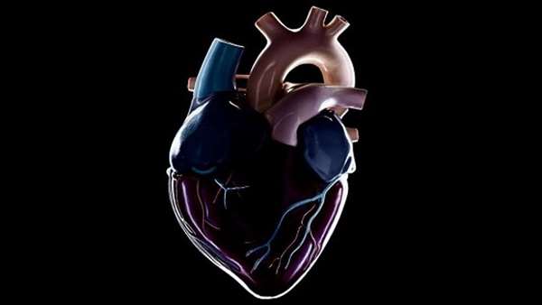
Stem Cells Renew Heart Function in Primates with Cardiac Failure
Researchers at the University of Washington, Seattle, have used human embryonic stem cell–derived cardiomyocytes (hESC-CMs) to restore heart function in macaque monkeys with heart failure.
Researchers at the University of Washington, Seattle, have used human embryonic stem cell–derived cardiomyocytes (hESC-CMs) to restore heart function in macaque monkeys with heart failure. The researchers hope that their achievement could pave the way for development of a similar treatment for human patients.
The macaque studies showed that the transplanted hESC-CMs developed into ventricular myocytes and formed electromechanical junctions with the animals’ own heart tissue, resulting in significant improvements in left ventricular function, and so the ability of the heart to pump blood. “In some animals, the cells returned the hearts’ functioning to better than 90% of normal,” comments Charles Murry, M.D., professor of pathology at the University of Washington School of Medicine, who headed the research team, which included collaborating scientists in the U.S., Ireland, Russia, and Taiwan. "This should give hope to people with heart disease.” Dr. Murry is also a professor of medicine in the division of cardiology, and of bioengineering, and is director of the UW Medicine Institute for Stem Cell and Regenerative Medicine.
The researchers report on their work in Nature Biotechnology, in a paper entitled “Human Embryonic Stem Cell–Derived Cardiomyocytes Restore Function in Infarcted Hearts of Non-Human Primates.”
Heart disease is the number one cause of death worldwide, at least in part because the heart has very limited ability to regenerate. After a heart attack, the damaged areas of heart muscle are replaced with scar tissue, which has no contractile ability, so the heart isn’t able to pump as efficiently and weakens. When the heart is no longer able to pump enough blood to supply the body with oxygen, the result is heart failure, which affects about 6.5 million people in the U.S., and causes more than 600,000 deaths every year. Current drug treatments can manage symptoms, “but do not address the root problem of muscle deficiency,” the authors point out
Over the last 20 years there has been an increasing focus on the development of cell-based therapies to promote heart regeneration, including the use of human cardiomyocytes derived from hESCs. Prior studies have shown that hESC-derived cardiomyocytes can survive and form new myocardial tissue that improves cardiac function in animal models of myocardial infarction.
For their latest studies, the authors evaluated intracardiac transplants of hESC-CMs in the macaque, a primate that represents a physiologically relevant large animal model. “This model should yield the best possible prediction of the human response to hESC-CM transplantation,” the team suggests. “The central hypothesis of this study was that hESC-CM would remuscularize the hearts of macaque monkeys and restore their function after myocardial infarction.”
The researchers induced experimental heart attacks in the monkeys, which after two weeks had reduced heart left ventricular ejection fraction (LVEF)—a measure of how well the heart is pumping—to about 40% of normal. This was enough to put the animals into heart failure.
At two weeks post infarction, the animals were then given injections of the hESC-CMs directly into the damaged areas of the heart and the surrounding heart tissue. The heart function was then followed using techniques including magnetic resonance imaging (MRI). The results showed that while LVEF in animals receiving sham injections steadily decreased even further over the next three months, LVEF in animals receiving hESC-CMs improved by an average of 10.6% after one month, and by three months there was an additional 12.4% improvement. The ejection fractions of two treated animals followed for three months continued to rise from 51% at four weeks after treatment, to 61% and 66%—essentially normal ejection fractions—at the three-month point. “The 20% absolute improvement in LVEF seen in the two hESC-CM hearts studied at 3 months was striking and shows that substantial mechanical improvement can occur between 1 and 3 months," the authors state.
Grafts in the treated animals averaged about 11% of the infarct size, and were shown to form electromechanical junctions with the host heart. By 3 months, the grafted tissue contained about 99% ventricular myocytes, the authors state. MRI scans in addition confirmed that new heart muscle had grown within the scar tissue in the treated hearts.
Previous research in monkeys has suggested that hESC-CMs can cause ventricular arrhythmias, and one of the animals in the University of Washington study also developed arrhythmias that “likely were due to the hESC-CM graft,” they note. However, the overall incidence of arrhythmias was lower than in previous studies with smaller infarcts, and the results of a separate, small infarct study the team carried out seemed to support this, “providing evidence that the lower burden of arrhythmias in this study resulted from the larger infarcts.”
Further studies indicated that the arrhythmias in hESC-CM animals were due to abnormal pulse generation and provided insights that the team say could help to identify either “pharmacological or cell-autonomous interventions” that could limit the likelihood of arrhythmias.
Encouragingly, functional recovery observed in the treated animals was larger than that observed in previous studies in rat or guinea pig models of myocardial infarction, even though the grafts were proportionally smaller. “This finding is likely explained by the greater physiological match between human and macaque,” the authors state. “The therapeutic effect may be further augmented when human cardiomyocytes are transplanted into diseased human hearts.”
"Our findings show that human embryonic stem cell–derived cardiomyocytes can remuscularize infarcts in macaque monkey hearts and, in doing so, reduce scar size and restore a significant amount of heart function," Dr. Murry comments. The team aims to develop a cell-based treatment that could be given shortly after a heart attack to prevent failure. Ultimately it may be possible to genetically alter the stem cell-based therapy to reduce the need for immunosuppression, and repeated treatments should be unnecessary because the cells are long lived. “What we hope to do is create a ‘one-and-done’ treatment with frozen ‘off-the-shelf’ cells that, like O-negative blood, can go into any recipient with only moderate immune suppression,” he suggests.
Reference:https://www.nature.com/articles/nbt.4162?_ga=2.250530886.1766938125.1530957027-741290648.1504594119


ارسال به دوستان