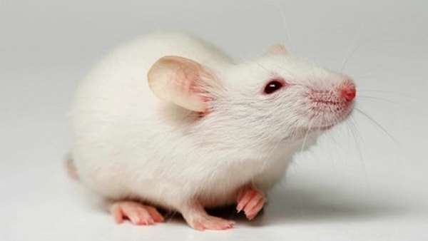
Cambridge scientists create first self-developing embryo from stem cells
Artificial mouse cells grown from outside body in a blob of gel shown to morph into primitive embryos, roughly equivalent to one third of way through pregnancy
The transformation of a fertilised egg into a tiny living embryo ranks among nature’s most impressive feats. Now scientists have replicated this critical step towards a new life for the first time, growing an artificial mouse embryo from stem cells in the lab.
The cells, grown outside the body in a blob of gel, were shown to morph into primitive embryos that perfectly replicated the internal structures that emerge during normal development in the womb.
The scientists let the artificial embryos develop in culture for seven days – about one third of the way through the mouse pregnancy. By this point the cells had organised into two anatomical sections that would normally go on to form the placenta and the embryonic mouse.
How scientists replicated a living mouse embryo using stem cells
Magdalena Zernicka-Goetz, a developmental biologist who led the work at the University of Cambridge, said: “I’m looking at it as a miracle of nature as well as trying to understand the process. It’s incredibly beautiful that we can begin to understand those forces that give rise to self-organisation during the earliest stage of development.”
The scientists’ goal is not to grow mice – or babies – outside of the womb. Instead, they are opening a new window on the embryo’s development just prior to implantation.
Until now scientists have struggled to recreate the embryo’s emerging three-dimensional structure outside of the body, while in the mother’s womb it is still too tiny to observe in detail using ultrasound.
“This is the time of implantation when the embryo is invading the body of the mother,” said Zernicka-Goetz. “Weeks later you can observe it with ultrasound but at this stage it is very mysterious. It’s a developmental black box.”
Scientists believe that as many as two-thirds of miscarriages take place before the embryo has implanted – and often before the woman is even aware that she is pregnant.
“To really understand the key principles of pregnancy at this stage would be very helpful,” said Zernicka-Goetz.
The team seeded the embryos from embryonic stem cells – powerful master cells that can turn into any cell type in the body – rather than starting from a fertilised egg, which could also potentially overcome the shortage of human embryos available for research. Currently, these are developed from eggs donated through IVF clinics, while the supply of embryonic stem cells is limitless.
“We are very optimistic that this will allow us to study key events of this critical stage of human development without actually having to work on embryos,” said Zernicka-Goetz. “Knowing how development normally occurs will allow us to understand why it so often goes wrong.”
Nick Macklon, professor of obstetrics and gynaecology at the University of Southampton, agreed that the research could pave the way to new insights that would help reduce miscarriage rates in the future. “If they are able to achieve this with human embryos I’m sure we will learn more about why early embryo development goes wrong and why some don’t implant,” he said. “This is … very impressive and important work.”
In the latest paper, published in the journal Science, single embryonic stem cells were mixed with small clusters of trophoblasts, the cells that go on to form the placenta.
The cells were placed in a semi-solid gel which allowed the structure to grow in three dimensions. After five days, the jumble of cells had multiplied and self-organised into distinct cell populations. The embryonic cells had also begun to diverge into two populations. One cluster, the mesoderm, would normally give rise to the heart, bones and muscles, while another contained cells that would go on to become brain, skin and eyes.
“It has anatomically correct regions that develop in the right place and at the right time,” said Zernicka-Goetz. “This was the most amazing thing for us.”
While the artificial embryo closely resembled the real thing, the researchers said it is unlikely that it would develop further into a healthy foetus. This would require the addition of the yolk sac, which provides nourishment for the embryo and within which a network of blood vessel develops.
The Cambridge team is now hoping to create similar artificial embryos with human cells.
Prof Robin Lovell-Badge, a stem cell biologist at The Francis Crick Institute in London, said one challenge was that scientists have not yet worked out how to extract trophoblast cells from human embryos.
In the mouse version, communication between the two cell types appeared to be what triggered the embryo to self-organise.
But, he added, “Scientists like to have unanswered questions – they keep us busy!”


ارسال به دوستان