
3D-printed Biomesh developed to promote hernia repair
Researchers from the Baylor College of Medicine (TX, USA) have developed a novel Biomesh to overcome postsurgical hernia complications. Their novel regenerative approach provides mechanical support to the injury site, whilst also acting as an inflammation modulating system.
Researchers from the Baylor College of Medicine (TX, USA) have developed a novel Biomesh to overcome postsurgical hernia complications. Their novel regenerative approach provides mechanical support to the injury site, whilst also acting as an inflammation modulating system.
Their research was recently published in Advanced Materials.
A hernia is a condition where an internal part of the body pushes through a weakness in the muscle or surrounding tissue wall. Most methods to repair a hernia consist of surgically implanting a prosthetic mesh to support and reinforce the damaged area. However, these mesh implants can adhere to the patient’s visceral organs – leading to chronic pain, bowel obstruction and bleeding as the mesh shrinks and hardens.
“Although hernia mesh implants are mechanically strong and support abdominal tissue, making the patient feel comfortable initially, it is a common problem that about three days after surgery the implant can drive inflammation that in two to three weeks will affect organs nearby,” commented lead author of the study, Crystal Shin (Baylor College of Medicine).
Inflammation can be controlled with medication; however these drugs may prevent the healing process by blocking the migration of immune cells to the injury site.
“To address these complications, we developed a non-pharmacological approach by designing a novel mesh that, in addition to providing mechanical support to the injury site, also acts as an inflammation modulating system,” Shin explained.
Inflammatory mediators known as cytokines accumulate at the mesh site after surgery. Many of these cytokines possess a property known as a positive surface charge.
“We hypothesized that Biomesh with a negative surface charge would capture the positively charged cytokines, as opposite electrical charges are attracted to each other,” explained study author, Ghanashyam Acharya (Baylor College of Medicine). “We expected that trapping the cytokines in the mesh would reduce their inflammatory effect and improve hernia repair and the healing process.”
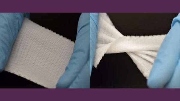
In their study, the team 3D printed a phosphate crosslinked poly (vinyl alcohol) polymer (X-PVA) Biomesh – and optimized this to withstand maximal abdominal pressure.
Remarkably, the team’s hypothesis was confirmed – the novel material was able to capture positively charged cytokines, all whilst retaining its elastic properties and remaining non-toxic to human cells.
In an animal study, the Biomesh was found to capture roughly three-times the amount of cytokines captured by the standard mesh. What’s more, the method effectively minimized postsurgical complications of hernia repair, with no visceral tissues adhering to the newly designed Biomesh.
“This Biomesh is unique and designed to improve outcomes and reduce acute and long-term complications and symptoms associated with hernia repair. With more than 400,000 hernia repair surgeries conducted every year in the [USA], the new Biomesh would fulfil a major unmet need,” Shin concluded.
“There is no such multifunctional composite surgical mesh available, and development of a broadly applicable Biomesh would be a major advancement in the surgical repair of hernia and other soft tissue defects. We are conducting further preclinical studies before our approach can be translated to the clinic. Fabricating the Biomesh is highly reproducible, scalable and modifiable.”
Link: https://www.regmednet.com/3d-printed-biomesh-developed-to-promote-hernia-repair/
ارسال به دوستان