Team uses organoid technology and single-cell RNA sequencing to study bladder cancer
Investigation of biological mechanisms in urothelial tissue, one of the three types of tissue that make up the urinary system, has always been very limited due to the lack of technologies that allow working with stable and long-term populations of the cells that compose it.
Investigation of biological mechanisms in urothelial tissue, one of the three types of tissue that make up the urinary system, has always been very limited due to the lack of technologies that allow working with stable and long-term populations of the cells that compose it. As a consequence, the mechanisms underlying diseases such as bladder cancer or cystitis remain poorly understood.
Now, scientists from the Epithelial Carcinogenesis Group led by Paco Real at the Spanish National Cancer Research Centre (CNIO) have used innovative organoid technology to achieve stable, long-term cultures of mouse urothelial cells. Subsequently, they characterised the organoids by sequencing the RNA of individual cells and identified a small population of stem cells—proliferative cells—that gives rise to the organoids and that could not be studied until now. This will allow working with this population and usher in a new era in the study of diseases of the urinary system. Researcher Catarina P. Santos is the first author of this study, which will be published in the journal Nature Communications.
First in-depth understanding of urothelial cells
Urothelial tissue is one of the three tissue types that make up the organs of the urinary system; it forms a layer that lines the bladder, kidneys, ureters and urethra. These cells are of great importance in the development of bladder cancer since their uncontrolled proliferation causes 90 percent of these tumours. These cells also play a role in other diseases, such as cystitis, which is very common, especially in women.
The in vitro study of these tissues has been hampered by the impossibility of achieving stable long-term cultures. "Until now, you could only keep the cells in culture for a few days or weeks, and you could hardly work with them because the cells quickly lost their personality," explains Real. "With this new technique, we were able to keep the cells in culture for more than a year, and the cells retain the properties of the original tissue."
Organoid technology allows cell cultures to grow in a three-dimensional matrix called Matrigel. Its advantage over traditional cultures in Petri dishes, which are two-dimensional, is that the three-dimensional structure is more similar to the conditions in which cells grow in the body itself. This technology could boost the generation of organs for transplantation in the future.
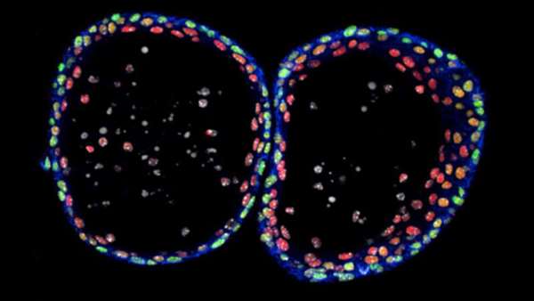
Molecular analysis of normal mouse urothelial organoids using immunofluorescence. Credit: CNIO
Investigation of biological mechanisms in urothelial tissue, one of the three types of tissue that make up the urinary system, has always been very limited due to the lack of technologies that allow working with stable and long-term populations of the cells that compose it. As a consequence, the mechanisms underlying diseases such as bladder cancer or cystitis remain poorly understood.
Now, scientists from the Epithelial Carcinogenesis Group led by Paco Real at the Spanish National Cancer Research Centre (CNIO) have used innovative organoid technology to achieve stable, long-term cultures of mouse urothelial cells. Subsequently, they characterised the organoids by sequencing the RNA of individual cells and identified a small population of stem cells—proliferative cells—that gives rise to the organoids and that could not be studied until now. This will allow working with this population and usher in a new era in the study of diseases of the urinary system. Researcher Catarina P. Santos is the first author of this study, which will be published in the journal Nature Communications.
First in-depth understanding of urothelial cells
Urothelial tissue is one of the three tissue types that make up the organs of the urinary system; it forms a layer that lines the bladder, kidneys, ureters and urethra. These cells are of great importance in the development of bladder cancer since their uncontrolled proliferation causes 90 percent of these tumours. These cells also play a role in other diseases, such as cystitis, which is very common, especially in women.
The in vitro study of these tissues has been hampered by the impossibility of achieving stable long-term cultures. "Until now, you could only keep the cells in culture for a few days or weeks, and you could hardly work with them because the cells quickly lost their personality," explains Real. "With this new technique, we were able to keep the cells in culture for more than a year, and the cells retain the properties of the original tissue."
Organoid technology allows cell cultures to grow in a three-dimensional matrix called Matrigel. Its advantage over traditional cultures in Petri dishes, which are two-dimensional, is that the three-dimensional structure is more similar to the conditions in which cells grow in the body itself. This technology could boost the generation of organs for transplantation in the future.
Thanks to single-cell RNA sequencing, the researchers were able to identify the nature of the urothelial stem cell population of these organoids that can proliferate and give rise to cancer. Until now, it was not possible to expand, analyse and describe this cell population because it accounts for less than 1 percent of the total bladder tissue. This makes it very difficult for researchers to work with these cells, and consequently, to make progress in the study of diseases resulting from their incorrect functioning.
"Now, we can look for these cells directly because we have more information about their identity," says Real. "We can also see how they resemble and how they differ from tumour cells, because tumour cells also proliferate inappropriately and they may share some similarities. This will give us clues as to what changed in those cells that led to the formation of a tumour."
New impetus for research into diseases and tissue regeneration
The team also showed that the acquisition of functional properties is dependent on Notch, a gene that in bladder cancer functions as a tumour suppressor: Notch activation causes cells to differentiate, which is a normal function of the cell that serves as a protection mechanism against tumour growth. "We show for the first time that this route, which we had previously demonstrated to be altered in bladder cancer, plays a role in normal tissue and conditions its differentiation, and that this is probably the reason why it also plays a role in tumours," says Real. "When this pathway is inactivated, the cells cannot differentiate well, and would be more susceptible to developing cancer."
These findings are not only of benefit for bladder cancer research, but also for other bladder pathologies such as cystitis. "Our work enables researchers who are investigating other pathologies to have stable cell populations in large numbers to study these diseases. In addition, our technique for expanding cell populations can be important for tissue regeneration after surgery."
As next steps in this research, Paco Real aims to perform the same procedure on human cells, genetically manipulate them, and then induce damage to analyse their behaviour in pathological situations and unravel their repair mechanisms.
Reference:https://www.nature.com/articles/s41467-019-12307-1


ارسال به دوستان