NC Researchers Engineer Lab-Grown Liver Tissue To Win NASA Challenge
Within our bodies, cells survive by being chatty: They rely on a network of blood vessels to communicate with one another and receive vital nutrients.
Within our bodies, cells survive by being chatty: They rely on a network of blood vessels to communicate with one another and receive vital nutrients.
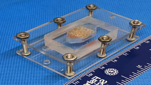
Replicating this cell crosstalk in a lab is less straightforward, but two teams of North Carolina scientists have done it, engineering lab-grown liver tissues that survived for 30 days. This breakthrough, which won the teams first and second prize in NASA’s Vascular Tissue Challenge, could lay the groundwork for better organ models and easier transplants in the future.
“When we opened the challenge back in 2016, we really weren’t sure if there would be a winner,” NASA Associate Administrator for Space Technology Jim Reuter said. “This is a really promising step for future tissue engineering studies.”
NASA’s Vascular Tissue Challenge tasked research teams with creating vascular tissue, or groups of cells that have blood vessels, that could sustain itself in a lab.
Both teams that completed the challenge hailed from the Wake Forest Institute for Regenerative Medicine in Winston-Salem. They used 3D printing to create gel-like scaffolds that the liver cells could use to exchange nutrients and oxygen.
The first team to complete the challenge, Team Winston, will receive $300,000 to continue their research, as well as an opportunity to test their tissue aboard the International Space Station U.S. National Laboratory. The second-place team, Team WFIRM, will receive $100,000. Team Winston was led by Professor James Yoo, and Team WFIRM by Director Anthony Atala.
Recreating vascular tissue function in a laboratory has promising implications. With further study of this proof of concept, scientists could use the tissue to model organ function from a lab bench, design organs fit for transplantation, and even study how the body reacts to long-term spaceflight.
Live Tissue On A Lab Bench
Keeping cell tissue alive outside of the body is no small feat. Researchers needed to design a system for the cells to communicate and receive nutrients like they would in a normal liver.
While both teams used 3D printing as part of their plan of attack, their approaches differed: Team Winston created a porous structure with many linking channels to mimic blood vessels, and Team WFIRM held cells together with a bridge that supported nutrient flow.
“It’s like a jigsaw puzzle, putting all these things together to finally make it work,” said Atala. “Once you finally get there, there’s a sense of satisfaction that the team has.”
The goal of NASA’s challenge was to engineer a tissue that was more than one centimeter thick. Now that both teams have accomplished it, they hope to build bigger tissues to better model the function of larger organs. Designing larger cell tissue models that can sustain themselves in the lab is a crucial next step to someday creating vascularized tissues for organ transplants.
“A lot of what we are looking at for the future is to be able to bring these structures together, [and] start building on these principles to start creating larger tissue structures that someday can go into patients,” Atala said.
Looking To The Future
Team Winston will work with the ISS U.S. National Laboratory to adapt their winning concept to a space test. The test could be used to see how the human body reacts to being in space, or to grow cell tissues in zero-gravity space conditions. In the meantime, NASA still has a third-place prize of $100,000 to award to the next team that completes the challenge.
For Yoo, the group’s success was the culmination of decades of work on advancing the study of the human body, both in space and on earth.
“What we have been doing for the past 30 years is to develop therapies that would help and benefit patients,” Yoo said. “Through this NASA challenge, we were able to make a breakthrough … and that is tremendous for us.”


ارسال به دوستان